
 Perfusion Imaging
We have been actively pursuing a neuroimaging research program exploring the brain basis for a behaviorally demonstrated cognitive deficit that is not revealed via standard neuroradiological (structural, magnetization prepared rapid gradient echo (MPRAGE)) imaging techniques.
By using perfusion imaging, it has been demonstrated that slowed and/or reduced blood flow can result in ‘functional lesions’ with serious cognitive consequences, producing behavioral deficits that may have no obvious neural basis when examined via standard structural imaging techniques. This research is continuing with those participants who have sustained both left and right hemisphere neural trauma.
Functional Neuroimaging
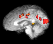
We have been using functional magnetic neuroimaging (FMRI) and evoked response potential (ERP) techniques as means of detailing the cerebral organization of language in both impaired and unimpaired populations. Research focuses on detailing the neural regions contributing to:
- the processing of various complex sentence types in hearing subjects ;
- the temporal parameters of speech;
- the effects of various methods standardly employed in language processing studies in brain recruitment and
We have investigated the role that standard psychological methodological tasks (probe verifications, thematic assignment, passive listening, etc.) have on inducing neural activation during comprehension with FMRI scanning. We have demonstrated a confound in the language and neuroscience literature. Namely, the effects purported to be caused by syntactic complexity of sentences is largely elicited only when certain, complex comprehension tasks were employed.